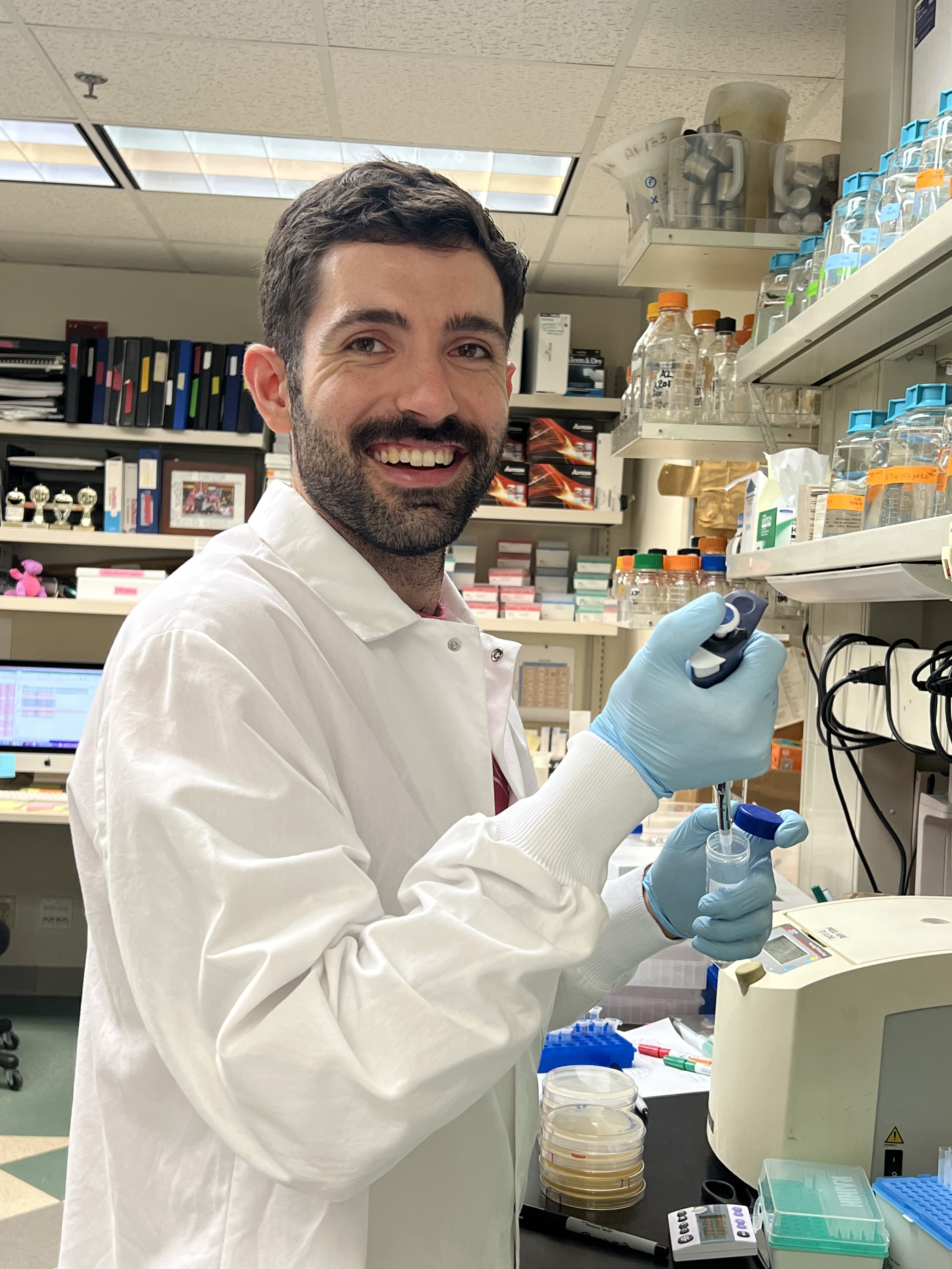
Like changes in key genes that control the cell cycle, changes to chromosomes can result in abnormal cell function and sometimes even cancer. Recently, a new type of genetic change has been linked to diverse cancers: the formation of circular DNA molecules from chromosomes. These molecules, known as extrachromosomal DNA or ecDNA, are dangerous because they do not follow the same rules of inheritance as normal chromosomes. Understanding the behavior of ecDNA within cells may uncover strategies to eliminate ecDNA and restore cellular health. Using a model ecDNA in budding yeast, Dr. King [HHMI Fellow] will identify and characterize pathways that either limit or enhance ecDNA propagation. He will then determine whether these pathways play a consistent role in human cancer cells, with the goal of identifying novel therapeutic vulnerabilities in treatment-resistant ecDNA-driven cancers. Dr. King received his PhD from the University of California, Berkeley and his BA from Columbia University, New York.
Human cells have complex mechanisms to repair DNA damage, such as that caused by exposure to sunlight or chemical substances. If DNA is not properly repaired, however, it can lead to cancer. In fact, faulty DNA repair has been associated with the initiation and progression of all types of cancer and is often targeted in cancer treatment to stop uncontrolled cell growth. A better understanding of how cells naturally defend against DNA damage will allow for the development of better drugs to treat cancer. Dr. Hoitsma [HHMI Fellow] aims to investigate specialized proteins, known as chromatin remodelers, that make damaged DNA accessible for repair. This research will provide insight for the development of novel therapeutic strategies to target these critical pathways. Dr. Hoitsma received her PhD from University of Kansas Medical Center, Kansas City and her BS from South Dakota State University, Brookings.
Neuroblastoma is a rare pediatric cancer that typically arises in the adrenal glands, located above the kidney. Children with high-risk neuroblastoma often have poor prognoses despite intense treatment-including maintenance treatment with retinoic acid-underscoring the need for new treatments to improve long-term outcomes. Retinoic acid, which is orally available and generally well tolerated, helps neuroblastoma cells mature (differentiate) into normal cells; however, this process is entirely reversible once the retinoic acid is withdrawn. If this differentiating effect could be made permanent with the addition of a second drug, a combination treatment with retinoic acid could become a novel method of preventing patient relapse. After testing a panel of 452 small molecule drugs, Dr. Weichert-Leahey discovered that a drug called PF-9363 accentuated the effects of retinoic acid in neuroblastoma the most. She will now study how PF-9363 functions, alone and together with retinoic acid, both in cells and patient-derived neuroblastoma models in mice. These experiments will indicate whether combinations of this new compound with retinoic acid may improve outcomes for children with high-risk neuroblastoma.
Courtney Ellison, PhD [Marilyn and Scott Urdang Breakthrough Scientist] is investigating how single bacterial cells join together to form complex, multicellular structures called biofilms. Biofilms protect bacterial cells from antibiotics and antimicrobial agents, making them difficult to eliminate. Some biofilm-forming species may cause certain cancers, and biofilms of infectious bacteria threaten immunocompromised patients such as those undergoing chemotherapy. Dr. Ellison focuses on bacterial appendages called type IV pili that play a crucial role in biofilm formation. Understanding the role of pili and their contribution to biofilm progression may lead to novel therapies to eliminate biofilms.

Dr. Andreeva focuses on structural and mechanistic aspects of inflammatory pathways underlying cancer and multiple other pathologies. Inflammation is an early response to various infections and damage initiated by the immune system. While it is essential for pathogen clearance and tissue repair, uncontrolled chronic inflammation promotes cancer development and all stages of tumorigenesis. Since inflammation is one of the hallmarks of cancer, understanding the processes leading to inflammation is critical for development of the next generation of cancer therapeutics. By using various structural, biochemical, and cell-based approaches, Dr. Andreeva aims to explain inflammatory pathways on a molecular level and provide a foundation for novel anti-inflammatory and anti-cancer treatments.
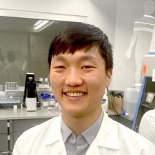
Cells in our body communicate with each other in a highly selective manner. These cell-cell interactions form the basis of numerous physiological functions, such as neuronal wiring and immune recognition. Dr. Shin plans to explore the general principles of cell-cell communication by constructing a synthetic synapse and studying its organization and functional diversity. His findings will elucidate the mechanisms that organize cell-cell interfaces involved in immune cell recognition of cancer and in the cell-type transitions associated with cancer and metastasis. This work will also provide a platform for engineering highly customized cell-cell interfaces, which may prove useful in engineering immune cell therapeutics.
This project employs the stickers-and-spacers model adapted from polymer physics. Macromolecules such as proteins and nucleic acids are described as a sequence of attractive domains called "stickers" and flexible, non-interacting domains called "spacers." Dr. Shin will use his lab's Monte Carlo simulation engine LaSSI (Lattice simulation engine for Sticker and Spacer Interactions) to calculate the average interactions between macromolecules and analyze their mesoscopic organization and phase properties.
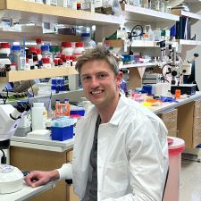
In both embryonic development and disease, the same genetic mutation can lead to highly variable outcomes in different individuals. Dr. Lammers aims to shed light on the drivers of this nongenetic variability using the developing zebrafish embryo as a model system. By combining fluorescence microscopy and single-cell sequencing, he will test whether subtle differences in gene expression within individual cells can explain why some embryos with a given genetic mutation survive to adulthood, while others perish within the first 24 hours of their development. His findings will provide a quantitative foundation for understanding the genetic and molecular basis of cancer outcomes in human patients where, for instance, tumors with the same underlying mutations often exhibit dramatically different disease courses.
Dr. Lammers will train Variational Autoencoders to learn low-dimensional latent space representations of whole-embryo transcriptomes and grayscale images depicting embryonic morphology. He will then train a third neural network to translate from transcriptional latent space to morphological latent space. Together, these three networks will comprise a new computational method, morphSeq, that takes single-cell transcriptomes of mutant and wildtype embryos as input and produces predictions for corresponding embryo morphologies as its output.
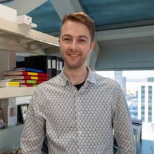
Cancer immunotherapy has revolutionized the way we treat cancer; however, it is only successful in a small subset of patients. Optimally functioning CD8 T cells, the specialized killers of the immune system, are key to the success of cancer immunotherapies. While CD8 T cell function is highly influenced by their metabolism, little is understood about how metabolism changes the function of these cells. Dr. Watson hypothesizes that metabolism affects CD8 T cell function by altering how tightly its DNA is packaged (its epigenetics), leading to altered gene expression. Using a mouse model of adoptive T cell therapy, a widely used immunotherapy in humans, and epigenetic techniques, Dr. Watson proposes to uncover how metabolism influences CD8 T cell epigenetic landscapes to control their function. He plans to apply these findings to improve T cell function and enhance tumor clearance. Dr. Watson received his PhD from the University of Pittsburgh, Pittsburgh and his BS from Hope College, Holland, Michigan.
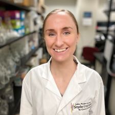
Neutrophils are important anti-microbial cells within the innate immune system. Recently, it has been shown that neutrophils can perform diverse functions, taking on both pro-inflammatory and pro-healing roles in response to tissue injury or insult. Dr. Siwicki's [Dale F. and Betty Ann Frey Fellow] goal is to understand how different neutrophil subtypes or states function to balance inflammatory versus regenerative processes, ultimately influencing tissue health and cancer. This work has the potential to uncover the basis of neutrophils' pro-tumor versus anti-tumor functions and could open the door to therapeutic targeting of specific neutrophil behaviors in order to improve clinical outcomes in cancer. Dr. Siwicki received her PhD from Harvard Medical School, Boston and ScB from Brown University, Providence.
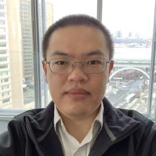
Global increases in metabolic syndrome, obesity, and diabetes are likely related to the overconsumption of hyper-palatable, cheap, ultra-processed food containing high amounts of added sugar and fat. Intriguingly, the vagus nerve has been discovered as the key conduit relaying information about sugar or fat ingestion from the gut to the brain, where a preference for sugar or fat is then developed and reinforced. Dr. Du [HHMI Fellow] aims to understand how the neurons are organized in the gut-brain vagal axis to sense sugar and fat, and to identify and characterize the neural circuits downstream of the gut-brain vagal axis that produce an insatiable appetite for sugar and fat. Understanding the basic biology of the gut-brain axis can provide important insights and strategies to help combat overconsumption of highly processed foods rich in sugar and fat, which may contribute to lowering the risk of metabolic diseases and cancer. Dr. Du received his PhD from The University of Texas Southwestern Medical Center, Dallas and his BS from the Tsinghua University, Beijing.
